Have you ever felt a sharp pain in your chest after a hard workout or a sudden fall? That pain might be coming from your costal cartilage. Most people have never heard of it. But this small piece of tissue does a big job inside your body. It connects your ribs to your breastbone. It helps you breathe, move, and stay protected. Understanding the anatomy of costal cartilage is the first step toward knowing why chest pain happens and how to handle it.
The costal cartilage is not bone. It is flexible tissue that bends with every breath you take. When it gets injured or inflamed, even simple movements can hurt. This guide breaks down exactly where it is, what it does, what causes pain, and how to treat and protect it in plain, simple language anyone can understand.
What Is Costal Cartilage?
Understanding the anatomy of costal cartilage is the semi-rigid connective tissue that links your ribs to your sternum (breastbone). It’s made of hyaline cartilage the same smooth, glassy tissue found in your nose tip and joint surfaces and it sits along the front (anterior) wall of the ribcage.
Unlike bone, costal cartilage is flexible. That flexibility isn’t a flaw; it’s by design. It allows your chest wall to expand and contract with every breath, absorb physical impact without shattering, and move naturally during exercise and daily activity.
All 12 pairs of ribs have costal cartilage at their front ends. However, the way each pair connects to the sternum varies significantly.
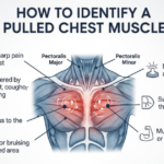
Related Post: How to Identify a Pulled Chest Muscle: A Complete Guide
Costal Cartilage Is Also Referred to As:
| Term | Meaning |
| Rib cartilage | Common lay term for costal cartilage |
| Hyaline rib cartilage | Refers to the specific type of cartilage tissue |
| Costal hyaline cartilage | Clinical anatomical term used in medical literature |
| Anterior rib extension | Describes its position at the front end of each rib |
Location of Costal Cartilage
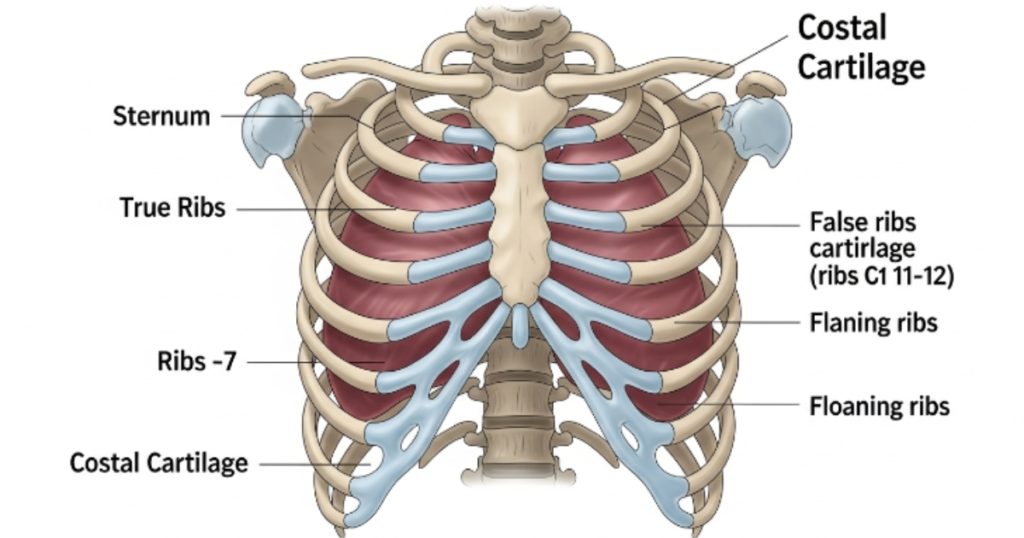
The costal cartilages are found exclusively along the anterior (front) surface of the thoracic cage, running between the bony rib ends and the lateral borders of the sternum. They are not present on the back or sides of the ribs.
Anatomical Placement of Costal Cartilage
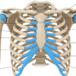
The 12 pairs of ribs are categorized into three groups based on how their cartilage connects or doesn’t to the sternum:
True Ribs (Ribs 1–7) Vertebrosternal Ribs Each of the first seven rib pairs has its own costal cartilage that attaches directly to the sternum. Ribs 1–7 are called “true ribs” because of this direct connection. They increase in length from rib 1 to rib 7, then gradually shorten.
False Ribs (Ribs 8–10) Vertebrochondral Ribs Ribs 8, 9, and 10 are called “false ribs.” Their costal cartilages don’t reach the sternum independently. Instead, each one connects to the cartilage of the rib directly above it. Together, they form the costal margin the curved boundary you can feel at the base of your ribcage.
Floating Ribs (Ribs 11–12) Vertebral Ribs The last two rib pairs have only small cartilaginous tips that don’t attach to anything. They float freely in the muscles of the lateral abdominal wall, hence the name. These ribs lack true costal cartilage connections.
Key anatomical note: Each costal cartilage is surrounded by a fibrous outer layer called the perichondrium, which provides structural strength and stiffness. As people age particularly women over 50 costal cartilages can undergo superficial ossification, gradually hardening and becoming more bone-like over time.
Costal Cartilage Function
Costal cartilage does far more than hold ribs to the sternum. It acts as the mechanical bridge that makes chest movement possible without constant fractures or joint damage.
Primary Functions of Costal Cartilage
1. Enables Breathing When you inhale, your diaphragm contracts and your ribcage expands. The costal cartilage bends slightly to accommodate this expansion, then recoils during exhalation. Without this flexibility, full lung inflation would be mechanically impossible.
2. Protects Vital Organs Along with the bony ribs, the costal cartilage completes the protective enclosure around the heart, lungs, and major blood vessels. It fills the gap between where bone ends and the sternum begins, leaving no unguarded space.
3. Shock Absorption During physical impact a fall, a sports collision, or blunt trauma costal cartilage absorbs and distributes force before it reaches internal organs. It acts as a natural bumper for the chest wall.
4. Supports the Costochondral Joint Where each rib meets its costal cartilage is called the costochondral joint. This junction allows subtle rotation and gliding movements that make upper-body flexibility possible, from twisting your torso to lifting heavy objects overhead.
5. Provides Chest Wall Stability The pectoralis major muscle attaches to the anterior surfaces of the upper six costal cartilages. The abdominal muscles attach to the lower cartilages. This means costal cartilage is literally a muscle anchor it plays a direct role in upper-body strength and movement.
Why Understanding the Costal Cartilage Is Important?
Most chest pain isn’t cardiac. Research consistently shows that a significant portion of patients presenting to emergency departments with chest pain are actually experiencing musculoskeletal chest wall issues many of which involve the costal cartilage.
Understanding this structure matters for several reasons:
- Accurate diagnosis: Costal cartilage injuries are often misidentified as muscle strains, heart issues, or even gastrointestinal problems. Knowing the anatomy helps narrow the cause faster.
- Better treatment decisions: Costal cartilage doesn’t heal the same way bone or muscle does. It has a limited blood supply, meaning injuries can take weeks to months to resolve.
- Preventing complications: Untreated inflammation like costochondritis can become chronic. Slipping rib syndrome caused by loose costal cartilage can lead to persistent, debilitating rib pain.
- Surgical relevance: Costal cartilage is commonly harvested for reconstructive procedures in rhinoplasty, ear reconstruction (microtia repair), and jaw surgery. Understanding its anatomy aids surgical planning and post-harvest recovery.
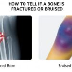
How To Know That Your Costal Cartilage Pain Is Serious?
Not all chest or rib pain requires an emergency room visit. But certain patterns should never be ignored. If your pain is sharp, persistent, or comes with other symptoms, it may signal a more serious condition.
Common Signs of Costal Cartilage Injury
| Symptom | What It May Indicate |
| Sharp chest pain at the sternum | Costochondritis or costochondral separation |
| Localized swelling over one rib | Tietze syndrome (2nd or 3rd rib most common) |
| Pain worsening on deep breathing | Costal cartilage inflammation or fracture |
| Tenderness on pressing the chest | Costochondral joint irritation |
| Pain radiating to the arm/shoulder | Possible cardiac cause seek emergency care |
| A clicking or “giving way” sensation | Slipping rib syndrome |
| Difficulty breathing or breathlessness | Requires immediate medical evaluation |
Go to the emergency room if you experience:
- Chest pain with sweating, nausea, or jaw pain
- Sudden severe chest pain after trauma
- Difficulty breathing that doesn’t resolve
- Pain that radiates into both arms
What Causes Costal Cartilage Pain?
Costal cartilage pain can develop from a variety of physical, inflammatory, and mechanical causes. It’s not always the result of a dramatic injury sometimes repetitive strain or even a prolonged cough is enough to trigger significant discomfort.
Causes of Costal Cartilage Pain
1. Costochondritis The most common cause of costal cartilage pain. It’s an inflammation of the cartilage at the junction between the ribs and sternum (the costochondral junction). Pain is usually sharp, located on the left side of the breastbone, and worsens with movement or pressure. It can affect multiple rib levels simultaneously and often has no clear cause.
2. Tietze Syndrome Similar to costochondritis but differentiated by visible or palpable swelling at the affected joint. It most commonly affects the 2nd or 3rd rib and tends to occur in people under 40. It’s considered a rare, benign, self-limiting condition that may follow respiratory illness with persistent coughing.
3. Direct Trauma A hard fall, car accident, or sporting impact can fracture or separate costal cartilage from the rib (costochondral separation). These injuries are often missed on standard X-rays and require CT imaging for diagnosis.
4. Repetitive Strain Heavy lifting, rowing, boxing, or any activity that repeatedly engages the pectoralis and intercostal muscles can irritate or strain the costal cartilage over time. This is common in athletes and manual workers.
5. Slipping Rib Syndrome Occurs when the costal cartilage connecting the false ribs (8–10) becomes loose, allowing the rib to slip and irritate nearby nerves. It causes intermittent, severe lower chest or upper abdominal pain.
6. Infection or Neoplasm Though rare, infection of the costochondral cartilage (infective chondritis) or cartilage destruction from tumors can present with localized pain, warmth, redness, and swelling. These require urgent medical investigation.
7. Arthritis and Systemic Inflammatory Conditions Conditions like rheumatoid arthritis, ankylosing spondylitis, and osteoarthritis can cause inflammation at the costochondral and sternocostal joints, mimicking or exacerbating costal cartilage pain.
How to Treat Costal Cartilage Injury?
Treatment depends on the underlying cause and severity. Most costal cartilage conditions are managed conservatively, but persistent or severe cases may require escalated intervention.
Rest and Activity Modification The first and most important step. Avoid any movement that aggravates the pain heavy lifting, twisting, or any exercise that puts pressure on the chest wall.
NSAIDs (Non-Steroidal Anti-Inflammatory Drugs) Over-the-counter medications like ibuprofen or naproxen help reduce both pain and inflammation. These are typically the first-line pharmaceutical treatment for costochondritis and Tietze syndrome.
Heat Therapy Applying a warm heating pad to the affected area can relax muscle tension around the ribcage and reduce aching. Several patients with Tietze syndrome report noticeable relief from consistent heat application.
Physical Therapy A physiotherapist can guide chest-wall stretching, postural correction, and breathing exercises. Studies show that structured stretching with a physiotherapist can meaningfully reduce pain in patients with symptoms lasting over a year.
Chiropractic and Osteopathic Care Spinal and thoracic alignment corrections may relieve pressure on the costal cartilage and improve mobility. Gentle rib mobilization can reduce costochondral joint stiffness.
Corticosteroid Injections For moderate to severe cases unresponsive to NSAIDs, a targeted injection of local anesthetic combined with a corticosteroid at the point of maximum tenderness can provide significant, lasting relief.
Surgical Intervention Rarely required. In extreme, persistent cases of Tietze syndrome or slipping rib syndrome, surgical resection of the affected cartilage may be considered. This is treated as a last resort and evaluated case by case.
Lifestyle Changes to Care About the Costal Cartilage
Long-term cartilage health doesn’t come from medication alone. Daily habits particularly exercise and diet play a significant role in maintaining strong, resilient costal cartilage.
Exercises for Costal Cartilage
A carefully structured exercise approach helps strengthen the muscles around the rib cage without straining the cartilage itself. Focus on low-impact, controlled movements:
- Diaphragmatic (belly) breathing: Trains proper breathing mechanics and gently mobilizes the costal cartilage without compressive force. Practice 5–10 minutes daily.
- Chest openers and gentle stretching: Standing in a doorway and gently pushing your arms back opens the anterior chest wall and reduces tightness in the costochondral region.
- Cat-Cow yoga pose: Improves thoracic spine mobility and reduces tension around the ribs and costal cartilage.
- Low-impact walking: Keeps the body active and promotes circulation to cartilage tissue without putting stress on the chest wall.
- Avoid: High-impact chest exercises, heavy bench pressing, or contact sports during acute or subacute costal cartilage pain phases.
Always consult a physiotherapist before starting exercises during an active injury phase. Incorrect loading can worsen costal cartilage inflammation.
Diet for Costal Cartilage
Cartilage has limited blood supply and heals slowly. Nutrition plays a vital supporting role in collagen synthesis, inflammation control, and tissue repair:
| Nutrient | Food Sources | Benefit for Cartilage |
| Vitamin C | Citrus fruits, bell peppers, broccoli | Essential for collagen production |
| Omega-3 Fatty Acids | Salmon, walnuts, flaxseeds | Reduces joint and cartilage inflammation |
| Collagen (or gelatin) | Bone broth, supplements | Provides raw material for cartilage repair |
| Calcium & Vitamin D | Dairy, eggs, sunlight, fortified foods | Supports bone and cartilage density |
| Antioxidants | Berries, leafy greens, turmeric | Reduces oxidative stress in connective tissue |
| Magnesium | Nuts, seeds, dark chocolate | Supports musculoskeletal function |
Foods and habits to avoid:
- Highly processed foods and refined sugar (increase systemic inflammation)
- Excessive alcohol (disrupts collagen synthesis)
- Smoking (reduces blood flow to cartilage tissue, impairing healing)
Final Verdict: Costal Cartilage Health Starts With Early Prevention
The costal cartilage is one of those structures you don’t think about until something goes wrong. And when it does, you feel it with every breath, every stretch, every movement of your upper body.
The good news is that most costal cartilage conditions including the most common ones like costochondritis are benign and respond well to conservative care. Rest, anti-inflammatories, physical therapy, and time are often all it takes to recover fully.
But waiting for pain to appear isn’t the right approach. Maintaining good posture, avoiding repetitive overloading of the chest wall, eating a collagen-supportive diet, and practicing proper breathing mechanics are all habits that protect your costal cartilage before problems start.
If you experience persistent chest pain, localized rib tenderness, swelling near the sternum, or pain that worsens with breathing, don’t dismiss it. Early diagnosis whether via physical examination, ultrasound, or CT imaging is always faster and cheaper than treating a condition that has been allowed to become chronic.
Conclusion
Costal cartilage is the hyaline cartilage that bridges your ribs and sternum, enabling every breath you take and protecting your vital organs from impact. When healthy, it works invisibly. When injured or inflamed, it can cause significant chest wall pain that disrupts daily life.
Understanding its anatomy from the true ribs (1–7) that connect directly to the sternum, to the false ribs (8–10) that join via shared cartilage, to the floating ribs (11–12) that attach to nothing helps both patients and clinicians identify the true source of thoracic discomfort.
Whether you’re managing an acute injury, recovering from costochondritis, or simply trying to keep your musculoskeletal system in good shape, the principles are the same: rest during flares, move with intention, eat to support collagen health, and seek professional guidance when symptoms persist beyond a few weeks.